Elbow Dislocation & Instability
Epidemiology & Mechanism
The elbow is the second most commonly dislocated major joint (after the shoulder), with an annual incidence of approximately 6 per 100,000. Simple dislocations (no associated fracture) are most common in young adults following sports injuries. Complex fracture-dislocations (terrible triad, Monteggia, Transolecranon) occur predominantly from higher-energy mechanisms in older patients.
Most (>90%) dislocations are posterolateral — the olecranon rotates and translates posterolaterally relative to the humerus, typically following a fall on an outstretched hand with the forearm in supination. The Hotchkiss ‘circle of injury’ describes the sequential disruption of stabilising structures from lateral to medial as energy increases: lateral collateral ligament complex → anterior and posterior capsule → medial collateral ligament complex.
Classification
Simple Dislocation | No associated fracture. Posterior or posterolateral. Usually treated with closed reduction and early mobilisation. Good prognosis. |
Terrible Triad | Elbow dislocation + radial head fracture + coronoid fracture. Highly unstable. Almost always requires surgery to stabilise the elbow and prevent recurrent dislocation. |
Radial Head Fracture-Dislocation (Essex-Lopresti) | Radial head fracture + elbow dislocation + interosseous membrane disruption. Results in proximal radial migration. |
Transolecranon Fracture-Dislocation | Olecranon fracture with anterior dislocation of the forearm. Requires ORIF. |
Monteggia Fracture-Dislocation | Proximal ulna fracture with radial head dislocation. Classified by Bado type. Requires surgical fixation. |
Emergency Management
- Closed reduction under conscious sedation or general anaesthesia: Gentle longitudinal traction with correction of medial/lateral displacement, followed by flexion of the elbow
- Post-reduction X-ray (AP and lateral): Confirms reduction and identifies fractures
- Stability assessment under fluoroscopy: Determines the functional arc of stability — if the elbow dislocates within 30–40° of full extension, surgical stabilisation is required
- Neurovascular examination: Anterior interosseous nerve (AIN) and ulnar nerve injuries occur in 10–20% of elbow dislocations
Treatment
Simple Dislocation — Stable After Reduction
- Sling for comfort for 1–2 weeks only — prolonged immobilisation causes stiffness
- Early range of motion: Supervised active elbow flexion-extension exercises from day 1 within the stable arc
- Physiotherapy: Restoring full motion and forearm rotation; return to sport at 4–6 weeks
Unstable / Complex Fracture-Dislocation (Surgical)
- Radial head fixation or replacement: A stable radial head is the primary lateral stabiliser — if fractured, it must be fixed (ORIF) or replaced with a metal implant (radial head arthroplasty)
- Coronoid fracture fixation: Even small coronoid fractures contribute critically to stability in the terrible triad — fixed with suture lasso technique or mini-screw
- LCL repair: The lateral collateral ligament complex is repaired to its humeral origin
- External fixation: Used as an adjunct when instability persists after all fractures and ligaments have been addressed
Chronic Elbow Instability
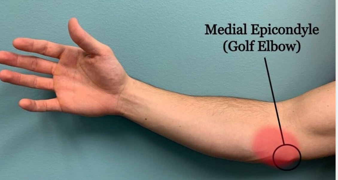
Posterolateral rotatory instability (PLRI) is the most common form of chronic elbow instability — caused by insufficiency of the lateral ulnar collateral ligament (LUCL) following a missed or inadequately treated dislocation. Patients report a sensation of the elbow ‘giving way’ or clunking, typically during weight-bearing on the outstretched arm. The lateral pivot-shift test (elbow flexion with supination and valgus stress under fluoroscopy) is confirmatory. Treatment: LUCL reconstruction using a palmaris longus or gracilis tendon graft.
Patient FAQs –Cubital Tunnel Syndrome (Ulnar Nerve Compression)
How quickly should an elbow dislocation be reduced?
As quickly as safely possible — ideally within 1–2 hours. Delayed reduction increases swelling, muscle spasm, and the risk of neurovascular compromise. If you or someone with you has an obvious elbow dislocation, go directly to the nearest A&E department. Do not attempt self-reduction.
Will my elbow ever be fully normal after dislocation?
Simple elbow dislocations in young patients have an excellent prognosis — >85% of patients regain a functional arc with no long-term instability. Loss of full terminal extension (5–10°) is common but rarely functionally significant. Complex fracture-dislocations have a more variable prognosis and may require longer rehabilitation.