Wrist Instability & Scapholunate Ligament Tears
Overview
Wrist instability describes a condition in which the carpal bones fail to maintain their normal interrelationships during loading and motion, due to ligamentous insufficiency. The scapholunate ligament — the primary stabiliser between the scaphoid and the lunate — is the most frequently injured intrinsic carpal ligament and the most common cause of wrist instability. An untreated complete scapholunate ligament tear leads inexorably to the SLAC wrist collapse pattern and end-stage wrist arthritis, typically within 10–25 years.
Mechanism & Presentation
- Mechanism: Fall on the outstretched hand with the wrist in extension and ulnar deviation — similar to a scaphoid fracture mechanism. The ligament may fail instead of (or alongside) the scaphoid bone.
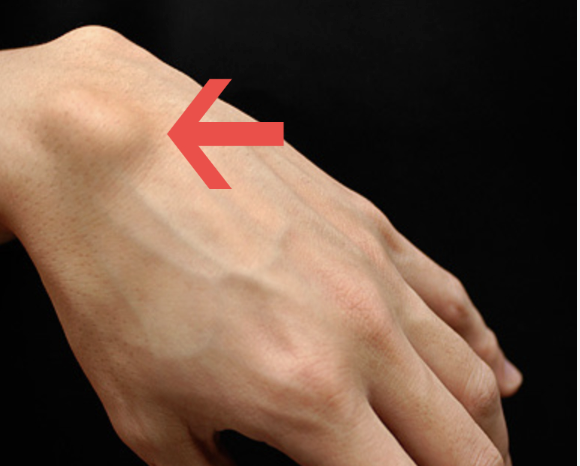
- Acute presentation: Dorsal wrist pain and swelling following a fall. X-ray may show a widened scapholunate gap (‘Terry Thomas sign’ — a gap >3mm between the scaphoid and lunate on AP view) or a rotatory deformity of the scaphoid (the ‘ring sign’ — foreshortening of the scaphoid on AP view from abnormal flexion).
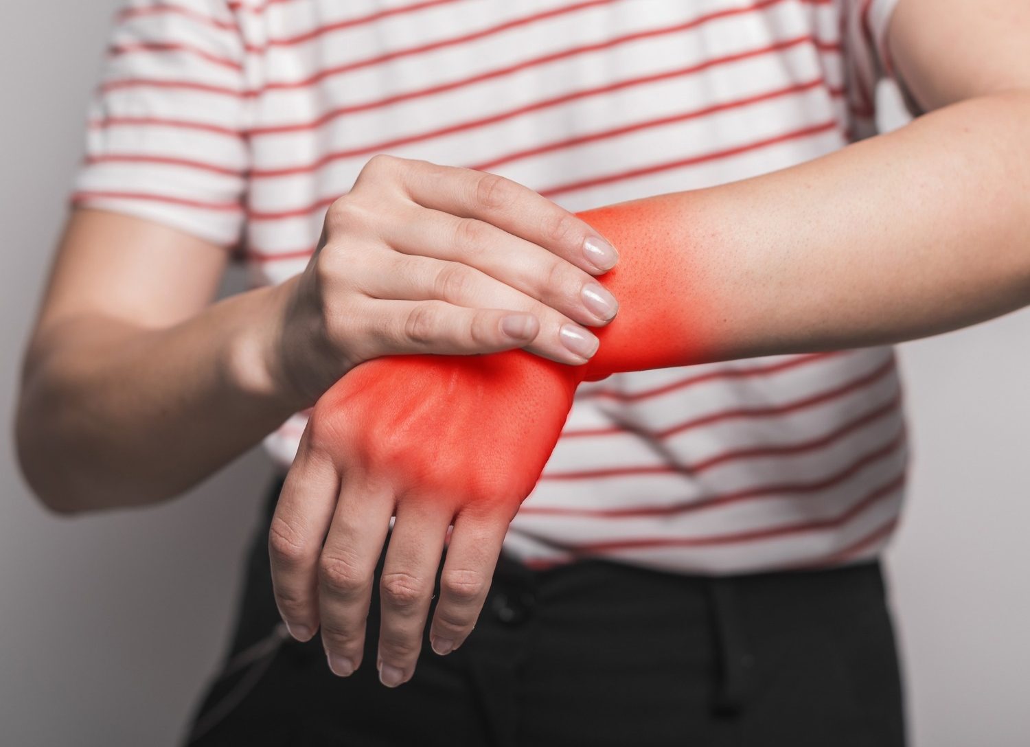
- Chronic presentation: Persistent dorsal wrist pain and weakness with gripping, a snapping or clunking sensation with wrist deviation, and progressive loss of wrist extension. The acute injury is frequently missed and presents months to years later as established instability.
Diagnosis
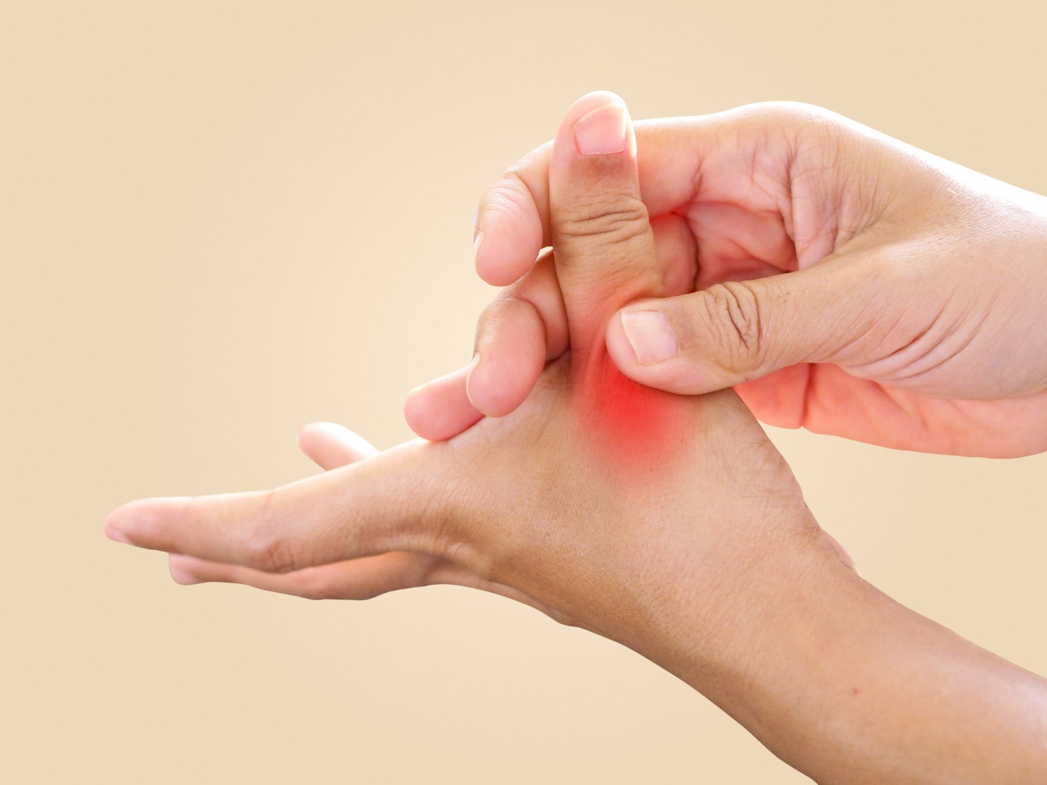
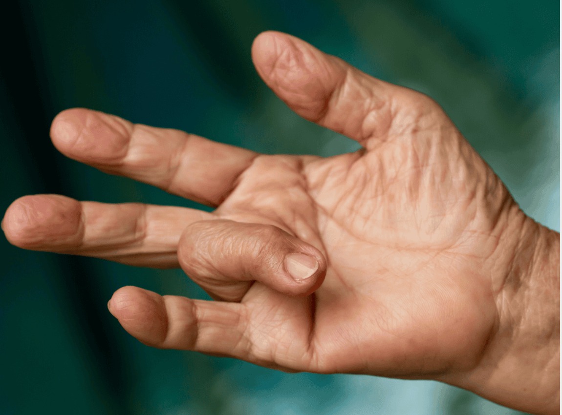
- Watson’s scaphoid shift test: The scaphoid is stabilised with thumb pressure on its tuberosity as the wrist is moved from ulnar to radial deviation — a painful clunk indicates scapholunate instability (sensitivity 69%, specificity 66%)
- X-ray: AP, lateral, and stress views. Key measurements: scapholunate angle (normal 30–60°; >70° = DISI deformity, pathological), scapholunate gap (>3mm abnormal on PA view with ulnar deviation and fist clenching)
- MRI arthrogram: Gadolinium contrast leaks through the torn ligament, confirming the diagnosis and grading the tear (Geissler arthroscopic classification I–IV)
Wrist arthroscopy: Definitive diagnostic tool and treatment platform — allows direct visualisation, probing, and grading of the ligament tear, and simultaneous repair or
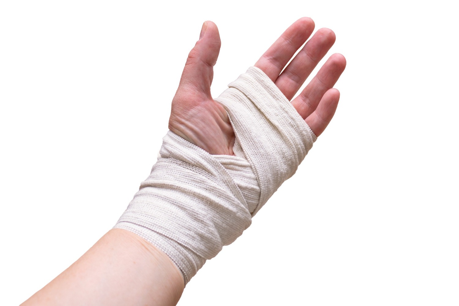
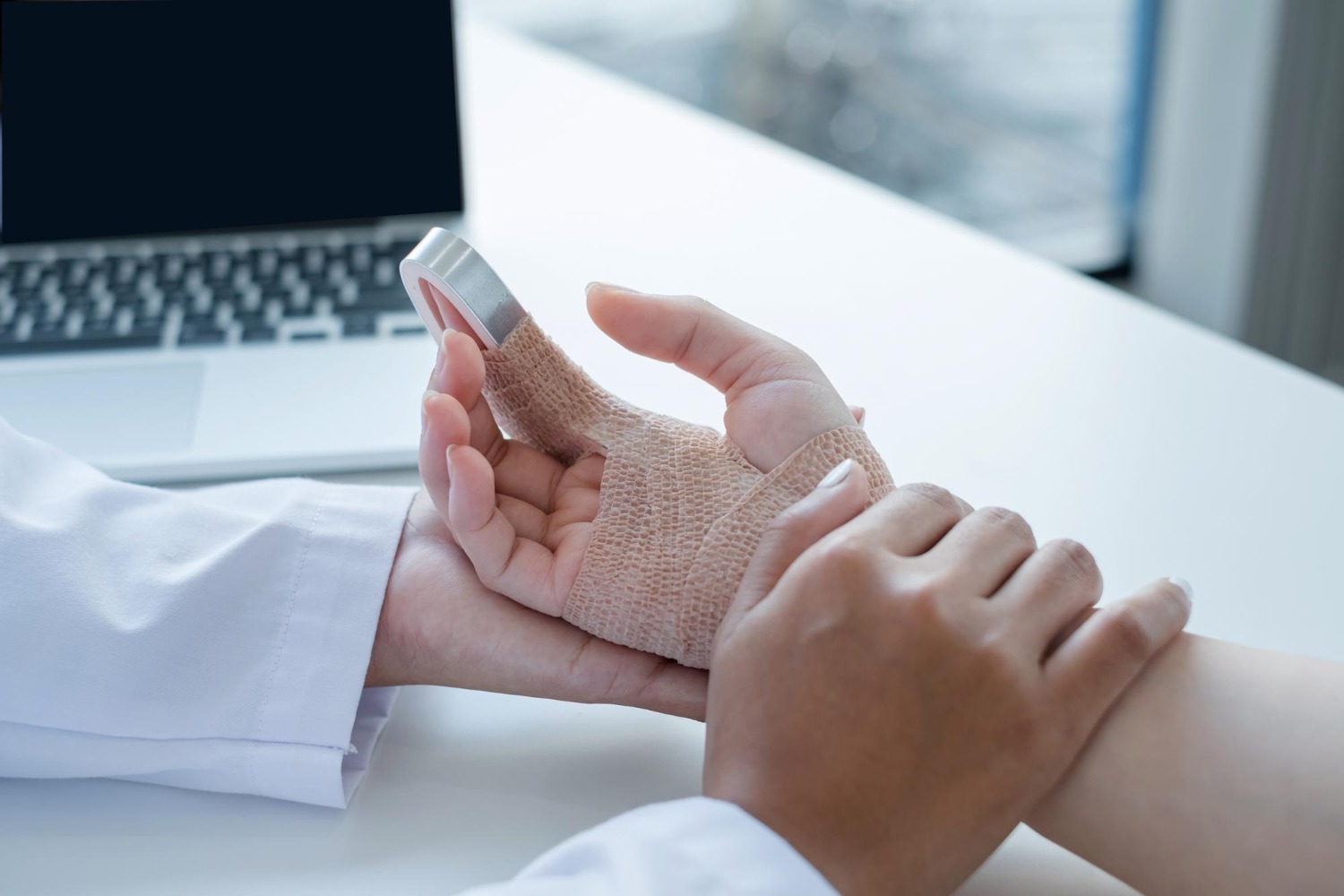
Treatment
Partial Tear (Grade I–II) | Cast immobilisation for 6–8 weeks; proprioceptive rehabilitation; arthroscopic thermal shrinkage or debridement for refractory cases |
Complete Tear, Acute (<6 weeks) | Arthroscopic or open primary repair — suture anchor reattachment of the ligament to the scaphoid dorsal margin, combined with temporary scapholunate K-wire stabilisation |
Complete Tear, Chronic (>6 weeks) | Primary repair not possible. Ligament reconstruction using tendon graft (modified Brunelli, RASL, or Corella techniques). Results less predictable than acute repair. |
With Early SLAC arthritis | Partial wrist fusion (STT fusion, scaphoid excision and 4-corner fusion) depending on extent of arthritis |
Patient FAQs –Wrist Instability & Scapholunate Ligament Tears
I was told I had a wrist sprain 6 months ago but it still hurts — could I have a ligament tear?
Yes. Scapholunate ligament injuries are frequently misdiagnosed as wrist sprains and undertreated. Persistent dorsal wrist pain, weakness with gripping, and a clunking sensation after a wrist injury should prompt specialist review. An MRI arthrogram can identify chronic ligament tears that plain MRI may miss. Prompt diagnosis significantly improves treatment options and outcomes.